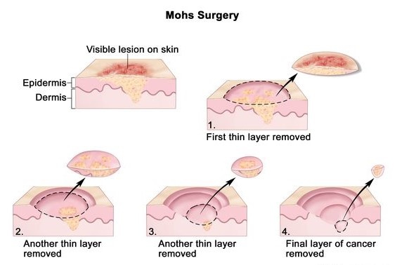
With Mohs micrographic surgery, the procedure is done in stages, including lab work, while the patient waits between each stage. After removing a layer of tissue, the surgeon examines it under a microscope in an on-site lab. If any cancer cells remain, the surgeon knows where they are and removes another layer of tissue from that location, while attempting to spare as much healthy tissue as possible. This process is repeated until no cancer cells remain.
| Date Taken: | 05.30.2023 |
| Date Posted: | 05.31.2023 16:42 |
| Photo ID: | 7828519 |
| VIRIN: | 230531-D-AB123-1001 |
| Resolution: | 565x378 |
| Size: | 59.96 KB |
| Location: | US |
| Web Views: | 34 |
| Downloads: | 4 |

This work, Murtha Cancer Center hosts annual Skin Cancer Summit [Image 2 of 2], must comply with the restrictions shown on https://www.dvidshub.net/about/copyright.