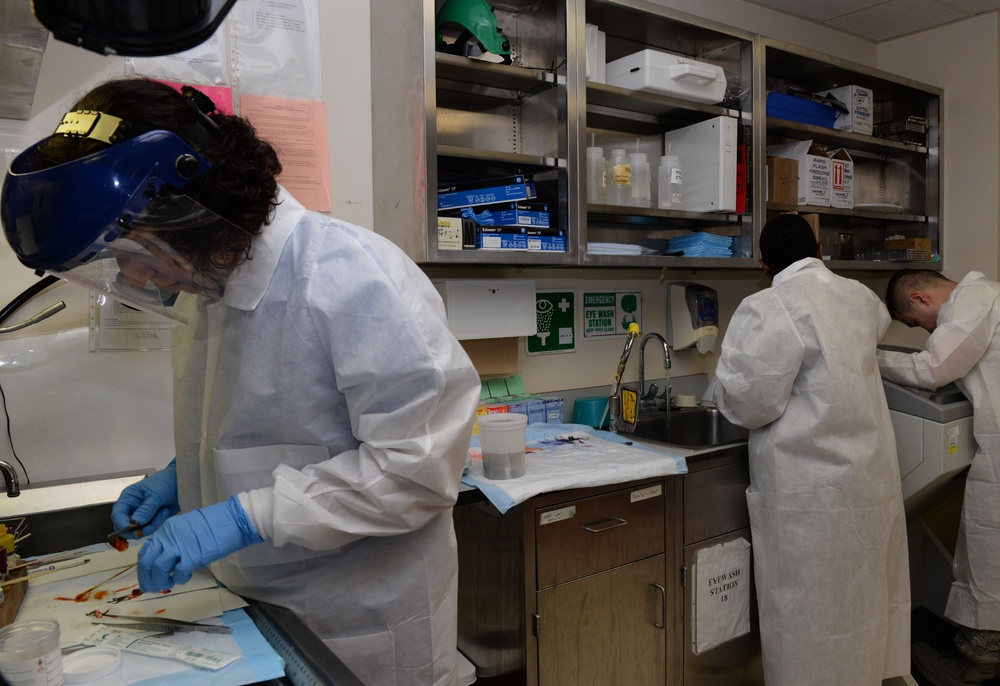
(left to right) U.S. Air Force Maj. (Dr.) Luisa Watts, 88th Diagnostics and Therapeutic Squadron pathologist, performs a gross prosection of an organ for intraoperative consultation (i.e. frozen section), while 88 DTS histopathology technicians, Airman 1st Class Simone Hairston and Staff Sgt. Steven Lam prepare the tissue samples selected by Dr. Watts for microscopic analysis inside the histology laboratory at Wright-Patterson Air Force Base Medical Center, June 26, 2017. Dr. Watts then reviews the resulting histology slide under a microscope to determine the type of surgery the patient will receive while the patient is still in the OR. (U.S. Air Force photo by Michelle Gigante/Released)
| Date Taken: | 06.25.2017 |
| Date Posted: | 07.19.2017 15:37 |
| Photo ID: | 3585293 |
| VIRIN: | 170626-F-AL359-1012 |
| Resolution: | 3000x2058 |
| Size: | 603.07 KB |
| Location: | OHIO, US |
| Web Views: | 34 |
| Downloads: | 6 |

This work, Wright-Patt histopathology laboratory diagnose disease through analyzing bodily fluids and tissues [Image 8 of 8], by Michelle Gigante, identified by DVIDS, must comply with the restrictions shown on https://www.dvidshub.net/about/copyright.