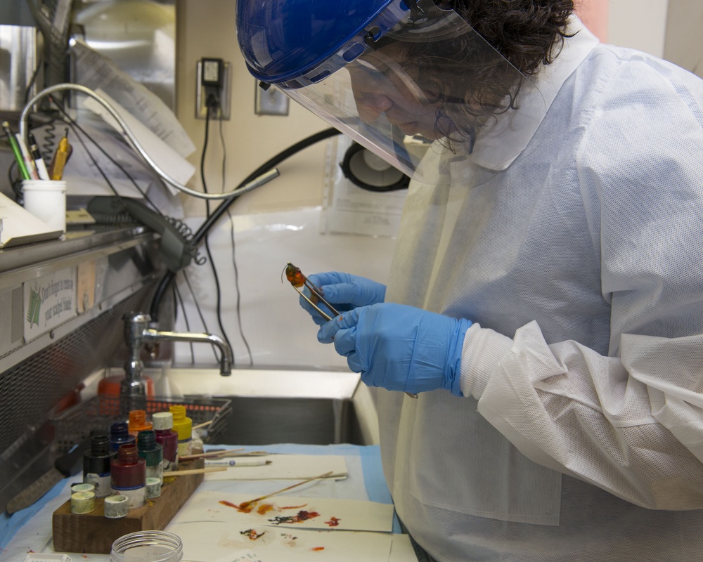
U.S. Air Force Maj. (Dr.) Luisa Watts, 88th Diagnostics and Therapeutic Squadron pathologist, inks a breast mass to maintain orientation for microscopic analysis inside the histopathology Medical Center at Wright-Patterson Air Force Base hospital, June 26, 2017. Dr. Watts will then cut the specimen into 3-5 mm slices, looking for abnormalities. She will choose which sections to have processed onto a histology slide to then review under the microscope before making her final diagnosis to determine the cause of the patient’s symptoms. (U.S. Air Force photo by Michelle Gigante/Released)
| Date Taken: | 06.25.2017 |
| Date Posted: | 07.19.2017 15:37 |
| Photo ID: | 3585513 |
| VIRIN: | 170626-F-AL359-1014 |
| Resolution: | 3000x2400 |
| Size: | 909.01 KB |
| Location: | OHIO, US |
| Web Views: | 59 |
| Downloads: | 8 |

This work, Wright-Patt histopathology laboratory diagnose disease through analyzing bodily fluids and tissues [Image 8 of 8], by Michelle Gigante, identified by DVIDS, must comply with the restrictions shown on https://www.dvidshub.net/about/copyright.